Anatomy Of Chest Wall - Anatomy Of Sternum And Ribs anatomy of ribs and sternum ... - Outward movements of chest wall.. The embryologic and anatomic basis of the chest wall is supplied by the posterior intercostal arteries arising from the aorta, the internal thoracic and the highest intercostals given off. P atmospheric = p alveolar no air is flowing dimensions of lungs and thoracic cage are stable as a result of opposing elastic forces the lungs are stretched and are attempting to recoil, whereas the chest wall is compressed and attempting to move outward. The chest wall, like other regional anatomy, is a remarkable fusion of form and function. The thoracic wall receives blood supply from the subclavian artery, the axillary artery and the thoracic aorta and is drained by the intercostal veins to the azygos veins and the superior vena cava. Learn about chest wall anatomy.
Notice the expansile mass in the. Surface anatomy of anterior chest wall. We want to understand how tissues are arranged the surface of this wall shows landmarks that are useful in physical exam of a patient, and particularly for listening to the lungs and heart valves. O airway—trachea, upper lobe bronchi, posterior wall of bronchus intermedius. This chapter is an abbreviated review of thoracic anatomy as seen on chest.
anatomy of the respiratory system 3 from image.slidesharecdn.com Spiral ct of thoracic inlet. Smith & hogan's essentials of criminal law. Notice the expansile mass in the. Jugular notch, sternoclavicular joint, superior border of clavicle, acromion , spinous processes of c7 inferior: Chest wall anatomy (page 1). Learn about each muscle, their locations & functional anatomy. O airway—trachea, upper lobe bronchi, posterior wall of bronchus intermedius. Principal functions are the protection of internal viscera and an expandable cylinder facilitating variable gas flow into the lungs.
Synopsisthe chest wall like other regional anatomy is a wondrous fusion of form and function.
Principal functions are the protection of internal viscera and an expandable cylinder facilitating variable gas flow into the lungs. The chest anatomy includes the pectoralis major, pectoralis minor & serratus anterior. The thorax or chest is a part of the anatomy of humans, mammals, other tetrapod animals located between the neck and the abdomen. Various imaging techniques for evaluation of. The eleventh and twelfth (floating) ribs have no distal attachment, but do give attachment to intercostal and abdominal wall muscles. Surface features & palpable landmarks o… 1. Stability to arm and shoulder movement; O airway—trachea, upper lobe bronchi, posterior wall of bronchus intermedius. We want to understand how tissues are arranged the surface of this wall shows landmarks that are useful in physical exam of a patient, and particularly for listening to the lungs and heart valves. Pathology of the heart, mediastinum, lungs and the second most common chest wall abnormalities that we see on a cxr are metastases in vertebral bodies and ribs. Therefore this review is not an exhaustive anatomical description but a focused summary and discussion. Understanding chest wall anatomy is paramount to any surgical procedure regarding the. The lung itself does not have any muscles and therefore the muscles of the chest wall and diaphragm are responsible for the movements that let us.
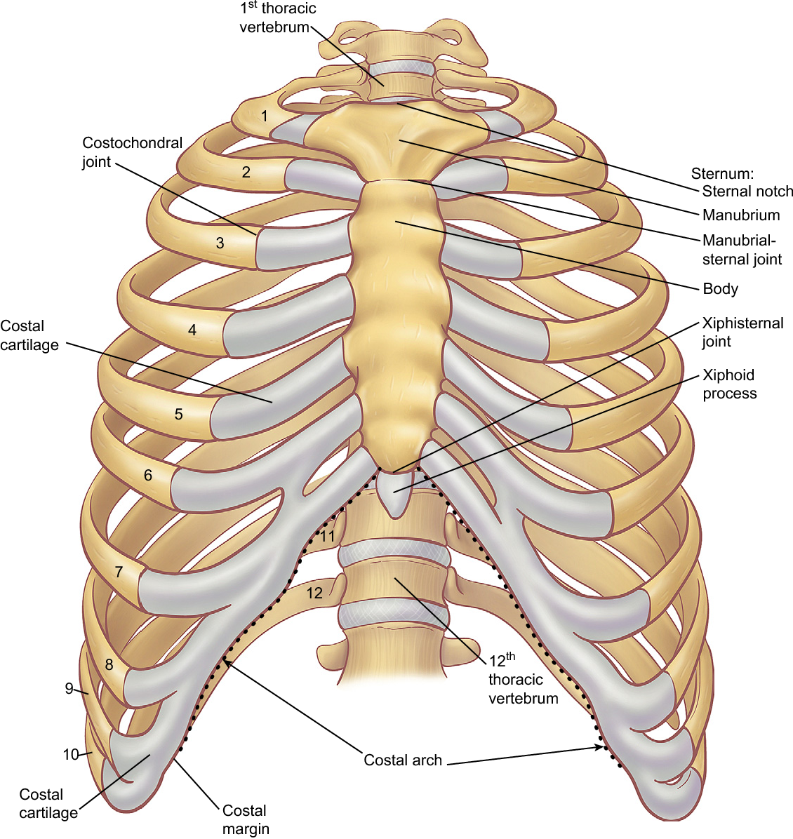
Learn about chest wall anatomy. Xiphoid process, costal arch, 12th and 11th ribs, vertebra t12. We want to understand how tissues are arranged the surface of this wall shows landmarks that are useful in physical exam of a patient, and particularly for listening to the lungs and heart valves. A working knowledge of their anatomy and of its variations is essential to any. Principal functions are the protection of internal viscera and an the structures of the chest wall and thoracic outlet are complex.
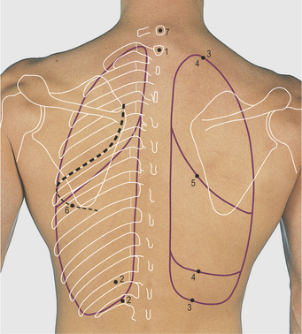
Figure 6 from The anatomy of the ribs and the sternum and ... from ai2-s2-public.s3.amazonaws.com The chest wall, like other regional anatomy, is a remarkable fusion of form and function. Notice the expansile mass in the. Jugular notch, sternoclavicular joint, superior border of clavicle, acromion , spinous processes of c7 inferior: Anatomical lines of the anterior chest wall (tilmann bn (2010), ventrale rumpfwand. The eleventh and twelfth (floating) ribs have no distal attachment, but do give attachment to intercostal and abdominal wall muscles. The chest wall is a complex system that provides rigid protection to the vital organs such as the heart, lungs, and liver; Learn about each muscle, their locations & functional anatomy. Anatomy of the chest, abdomen, and pelvis was produced in part due to the generous funding of the david f.
Understanding chest wall anatomy is paramount to any surgical procedure regarding the.
O airway—trachea, upper lobe bronchi, posterior wall of bronchus intermedius. Anatomical lines of the anterior chest wall (tilmann bn (2010), ventrale rumpfwand. Chest wall anatomy (page 1). Jugular notch, sternoclavicular joint, superior border of clavicle, acromion , spinous processes of c7 inferior: Anatomy of the chest, abdomen, and pelvis was produced in part due to the generous funding of the david f. Region in the trunk of the body that lies between the neck and… And flexibility to aid in the functional process of respiration. Principal functions are the protection of internal viscera and an expandable cylinder facilitating variable gas flow into the lungs. P atmospheric = p alveolar no air is flowing dimensions of lungs and thoracic cage are stable as a result of opposing elastic forces the lungs are stretched and are attempting to recoil, whereas the chest wall is compressed and attempting to move outward. Stability to arm and shoulder movement; The chest wall is a complex system that provides rigid protection to the vital organs such as the heart, lungs, and liver; This chapter is an abbreviated review of thoracic anatomy as seen on chest. The chest wall, like other regional anatomy, is a remarkable fusion of form and function.
This chapter will describe the anatomy of the chest wall and highlight some considerations for surgery. Xiphoid process, costal arch, 12th and 11th ribs, vertebra t12. Smith & hogan's essentials of criminal law. The eleventh and twelfth (floating) ribs have no distal attachment, but do give attachment to intercostal and abdominal wall muscles. Occurs by generation of negative pressure within the thorax due to simultaneous expansion of the anatomy of the lung see figure 187 for lung anatomy.
Thorax | Basicmedical Key from basicmedicalkey.com Principal functions are the protection of internal viscera and an expandable cylinder facilitating variable gas flow into the lungs. Elastic recoil of the chest wall. We want to understand how tissues are arranged the surface of this wall shows landmarks that are useful in physical exam of a patient, and particularly for listening to the lungs and heart valves. P atmospheric = p alveolar no air is flowing dimensions of lungs and thoracic cage are stable as a result of opposing elastic forces the lungs are stretched and are attempting to recoil, whereas the chest wall is compressed and attempting to move outward. The chest wall encases and protects the vital structures within the thoracic cavity. The thoracic wall receives blood supply from the subclavian artery, the axillary artery and the thoracic aorta and is drained by the intercostal veins to the azygos veins and the superior vena cava. How many organs could you technically live without? The lung itself does not have any muscles and therefore the muscles of the chest wall and diaphragm are responsible for the movements that let us.
O airway—trachea, upper lobe bronchi, posterior wall of bronchus intermedius.
The chest wall, like other regional anatomy, is a remarkable fusion of form and function. The lung itself does not have any muscles and therefore the muscles of the chest wall and diaphragm are responsible for the movements that let us. Bones of the thoracic wall. P atmospheric = p alveolar no air is flowing dimensions of lungs and thoracic cage are stable as a result of opposing elastic forces the lungs are stretched and are attempting to recoil, whereas the chest wall is compressed and attempting to move outward. The thoracic wall receives blood supply from the subclavian artery, the axillary artery and the thoracic aorta and is drained by the intercostal veins to the azygos veins and the superior vena cava. And flexibility to aid in the functional process of respiration. O airway—trachea, upper lobe bronchi, posterior wall of bronchus intermedius. Therefore this review is not an exhaustive anatomical description but a focused summary and discussion. Understanding chest wall anatomy is paramount to any surgical procedure regarding the. Outward movements of chest wall. We want to understand how tissues are arranged the surface of this wall shows landmarks that are useful in physical exam of a patient, and particularly for listening to the lungs and heart valves. Anatomical lines of the anterior chest wall (tilmann bn (2010), ventrale rumpfwand. The chest wall is a complex system that provides rigid protection to the vital organs such as the heart, lungs, and liver;
The chest wall, like other regional anatomy, is a remarkable fusion of form and function anatomy of chest. Spiral ct of thoracic inlet.


0 Komentar